Departments join forces for zebra X-ray
The Department of Radiology teamed with University of Iowa Health Care Information Systems and the UI Museum of Natural History to get an X-ray of a taxidermic zebra to prepare to conserve the mounted animal this winter.
October 3, 2021
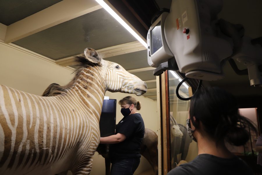
Three University of Iowa departments teamed up this past summer to get an X-ray of the taxidermic zebra displayed in the UI Museum of Natural History’s Mammal Hall.
The project was organized by the Carver College of Medicine’s Department of Radiology, UI Healthcare Information Systems, and the Museum of Natural History.
The nearly 100-year-old zebra was X-rayed to give Goldberg Preservation Services conservator Lisa Goldberg the information necessary to properly preserve the zebra when Goldberg arrives on campus this winter. The conservation project will address light and environmental degradation the zebra has experienced over time, according to the Pentacrest Museums website.
Jessica Smith, the communications coordinator for the Museum of Natural History, said the process started when the conservators asked if it would be possible to get an X-ray before beginning their work. Museum Director Liz Crooks contacted the UI Library to help with additional contacts.
“Through a long chain of resources, all within the University of Iowa, this partnership came to be,” she said.
Janet Roe, technical director at the Department of Radiology at UI Hospitals and Clinics, got involved when the Pentacrest Museums reached out to the radiology department about the project. From there, Roe said, it was a matter of figuring out logistics.
Because the zebra is fragile, having been around since before the 1930s, the Department of Radiology had to come to its exhibit, Smith said. The department borrowed a truck to transport the nearly one-ton portable X-ray used for the project. Roe said transporting it was tricky, as the machine would sometimes rock when the truck turned a corner.
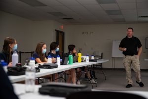
Once the X-ray arrived at the museum, radiologic technologists Rebecca Egbert, Alicia Waite, and Kelli Zimmerman were tasked with maneuvering the machine, getting angles to provide the most useful information to Goldberg, who observed over video chat.
The entire operation took place inside the zebra’s case to avoid damaging it.
“It really reinforced to me how valuable [radiologic technologists] are in providing health care,” Roe said. “Hospitals don’t run without radiologic technologists.”
The X-rays also helped Goldberg explore the historic taxidermy techniques used by Carl Akely, back in the beginning of the 20th century. Akely was a taxidermy pioneer, who developed a more precise technique for taxidermy that involved plaster and clay molds, in a method that would come to be known as the “Akely Method of Taxidermy.”
Roe said the radiologists found wood, screws, and foam inside the zebra that help it maintain its structural integrity. The zebra’s ears are held up with lead.
Smith said this kind of interdepartmental collaboration makes the UI special.
“We have a lot of different experts in a lot of different fields who are not stingy with their expertise at all,” she said. “This was an especially generous partnership on part of our colleagues at the Department of Radiology and we are incredibly grateful.”
Colin Derdeyn, chair of the Department of Radiology in the Department of Medicine, said collaborative work happens in other departments, citing work related to cystic fibrosis with UI Professor John Englehardt.
“Bringing together his model and our techniques, we’ve got a real opportunity to figure out some ways that cystic fibrosis causes problems that can be fixed,” Derdeyn said. “It’s [in] bringing together things like that where all the fun is.”
The zebra exhibit can be viewed in Mammal Hall when the museum is open from 10 a.m. to 5 p.m. on Fridays and Saturdays.