

UI Heart and Vascular Clinic completes 1000th TAVR surgery
The UI Heart and Vascular Center completed its 1,000th transcatheter aortic valve replacement procedure, an alternative surgery for high-risk patients that cannot undergo open heart surgery.
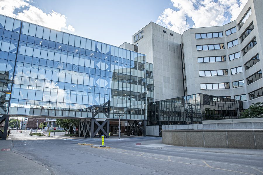
University of Iowa Hospitals and Clinics are seen on Tuesday, June 23, 2020.
July 14, 2020
Cardiac surgeons and cardiologists in the University of Iowa Heart and Vascular Center attribute their recent award as best in the region to the experience and collaborative nature of team members.
The UI Heart and Vascular Center recently performed its 1000th transcatheter aortic valve replacement (TAVR) procedure, which is an alternative procedure for high-risk patients who suffer from severe aortic stenosis – diseased valves – and cannot undergo open heart surgery.
UI Assistant Professor of Surgery Sharon Beth Larson, who works in the division of cardiothoracic surgery, said the transcatheter aortic valve replacement procedure was specifically targeted to patients in their late 80s and 90s before developments were made. She said they would benefit from valve replacement but were not quality surgical candidates.
“Every procedure has a risk for complications,” Larson said. “There are risks to open heart surgery, and we found in the early days of [this procedure] that there was a patient population where no surgeon would touch that patient, so we knew we needed other methods of [surgery].”
Open-heart surgery uses a bypass machine to cut into the diseased valve in the heart, which Larson said results in longer recovery and rehabilitation time. The transcatheter aortic valve replacement uses a catheter, much like a small metal straw, and inserts it into the seminal artery to load the new valve into the bloodstream and up to the heart.
RELATED: New UIHC procedure provides safer treatment options for high-risk heart patients
The surgeons then inflate a balloon-like structure that pushes the diseased valve into the heart’s tissue wall to make room for the new valve. Though there is a risk of bleeding or heart rupture, Larson said the procedure is minimally invasive and complications occur less than 1 percent of the time.
The procedural team is composed of cardiologists, cardiac surgeons, cardiac anesthesiologists, and imaging cardiologists. Each has their own role that contributes to a patient’s surgery and recovery, she said.
UI Clinical Professor of Internal Medicine Phillip Horwitz, co-manager of the team, said the transcatheter aortic valve replacement program has come a long way since its establishment in winter 2011.
“In 2011, we did our first procedure,” Horwitz said. “To now have 1,000 under our belt, it is exciting to be a part of a program like this and develop a new technology that has changed the way we take care of [severe aortic stenosis].”
He said surgical aortic valve replacement has been around for decades and was considered the gold standard of treatments, but after the UI Heart and Vascular Center began treating unlikely candidates for surgery, they were able to expand the technology to include medium and low-risk patients.
“We started with the sickest group of patients and now research has shown over the last decade that this technology can be used for patients with medium-risk complications,” Horwitz said. “It’s gone from working with the sickest to it now being routine for most patients.”
Larson said that surgeons and cardiologists are present for the surgery, along with a support staff to assist them, and cardiac anesthesiologists help to monitor the function of the heart. She said collaboration among team members with various specialties is what makes UI Hospitals and Clinics unique.
“We are the center for the most specialized care,” Larson said. “There is nothing we cannot take care of and we not only have the ability, but when resources and skill sets are exhausted in other hospitals have been exhausted, we are a beacon of hope where we help take care of them.”
UI Clinical Assistant Professor of Internal Medicine and cardiologist Sidakpal Panaich said it is vital to a patient’s health and safety that cardiologists and cardiac surgeons partner with imaging cardiologists to assess all areas of surgery.
He said cardiac imaging allows for surgeons to determine how to use the right surgical approach and target the correct valve to minimize risks. Imaging can also eliminate patients that clinically might be able to undergo surgery but through scans are determined to no longer be a candidate, he added.
“It’s a wholesome, collaborative approach from surgeons to imaging colleagues,” Panaich said. “It is our general cardiologists who we depend on for excellent post-procedural care. There are some delayed issues that need to be checked and managed.”
He said it was an amazing opportunity to not only be part of a great surgical team, but part of a great institution in general.
“We are able to provide cutting-edge procedures; we can provide resources for our patients,” Panaich said. “We start these newer programs early in their development, so we are able to offer these programs to other institutions and gain more and more experience. Anyone can put in a valve, but it is about the planning and having the right team and picking the right patient. Taking care of complications with a hard team approach and experience sets us apart.”